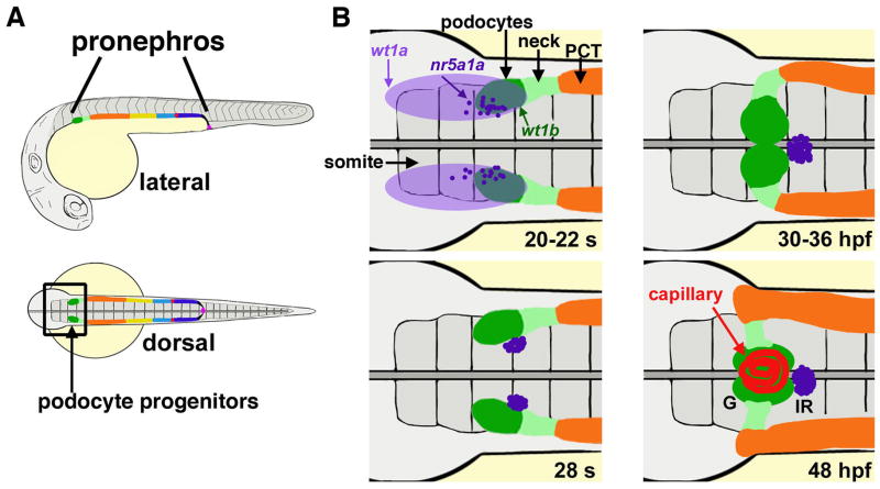
FIGURE 3. Zebrafish podocyte lineage specification and glomerular development.
(A) The zebrafish pronephros contains podocytes (dark green) at the rostral-most position. (B) Developmental timecourse of the cell populations that develop in proximity to podocytes. Gene expression of wt1a (light purple) is broad, while wt1b transcripts (dark green) are restricted next to somite (s) three, and interrenal precursors marked by nr5a1a transcripts (dark purple) are interspersed in this region. The neck (light green) is located caudal to the podocytes, followed by the proximal convoluted tubule (PCT, orange). Cell movements between the 20 somite stage to 48 hours post fertilization (hpf) lead to formation of a single glomerulus (G) with central capillary nexus (red). The interrenal gland (IR) (dark purple) is situated just caudal to the glomerulus. [Figure adapted from Wiley Interdiscip Rev Dev Biol, 2, Gerlach G, Wingert RA, Kidney organogenesis in the zebrafish: Insights into vertebrate nephrogenesis and regeneration, Pages 559–585, Copyright 2013, with author permission].

