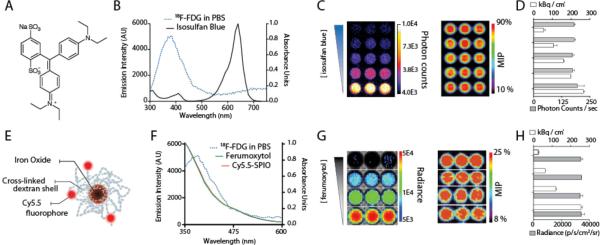
Figure 2.

Cerenkov Quenching with Clinical Applied Materials. (A) Chemical structure of the patent blue dye Lymphazurin, used for sentinel lymph node imaging. (B) CL profile and absorption spectra of [18F]-FDG and patent blue dye, respectively. (C,D) Dose-dependent quenching of CL signal by Lymphazurin; PET signal remains unaffected. (E) Schematic of iron oxide nanoparticle, modification with fluorescent dyes. (F) CL profile of [18F]-FDG and absorption spectra of the FDA-approved ferumoxytol and the research grade Cy5.5-SPIO. (G,H) Again, a concentration gradient of the ferumoxytol produces a change in the amount of the CL signal produced, while PET signal remains constant. MIP, mean intensity projection of the tomographic PET dataset. Photon counts are counts/sec; Radiance units are photons/cm2/sr/sec.
