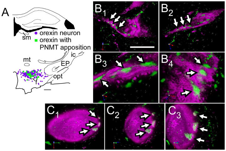
Figure 1.
Innervation of orexinergic neurons by C1 cells.
A, Computer-assisted drawing of PNMT contacts within orexin neurons.
B1–B4, C1–C3. Close appositions between PNMT terminals (revealed with Alexa-488, green fluorescence) and orexin (with Cy-3, in magenta) neurons in rats. Close appositions indicated by arrows. Putative synaptic contacts revealed through velocity 3D rendering software using deconvoluted images of 19 (in B) and 29 (in C) serial 0.3 μm Z-stacks. The overlapping region of the two fluorophores is seen as white pixels (visible in B3, B4 and C2, C3) and represents the putative contact zone.
Abbreviations: EP, entopeduncular nucleus; f, fornix; ic, internal capsule; mt, mammillothalamic tract; opt, optic tract; sm, stria medullaris of the thalamus
Scale bar A, 500 μm; B1–B2, 20 μm; B3–B4, 5 μm; C1–C3, 10 μm.

