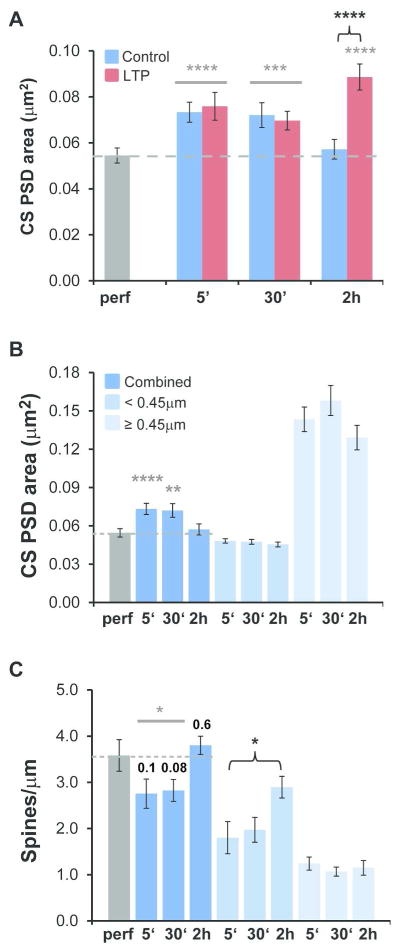
Figure 6.
Restoration of small dendritic spines and average PSD area in acute slices to levels found in perfusion-fixed hippocampus. (A) Relative to perfusion-fixed hippocampus (dashed gray line in each graph), PSD area was increased significantly in slices under both control and LTP conditions at 5 min (hnANOVA, F(1,432) = 19.2, p < 0.0001, gray****) and 30 min (hnANOVA, F(1,529) = 7.0, p < 0.001; gray***, but in only two out of three experiments). In the 2 hr LTP condition, PSD area was elevated relative to perfusion-fixed hippocampus (hnANOVA, F(2,498) = 19.4, p < 0.0001, post-hoc Tukey, p < 0.0001, gray****) and the 2 hr control condition (post-hoc Tukey p < 0.0001, black bracket****), while the 2 hr control condition was comparable to the perfusion-fixed condition (post-hoc Tukey, p = 0.97). (B) Restricting these comparisons to perfusion-fixed versus slice control conditions (data shown in A) revealed greater PSD areas (hnANOVA, F(3,932) = 10.4, p < 0.0001) at 5 min (post-hoc Tukey, p < 0.0001****) and at 30 min (p < 0.01**), but not at 2 hr (post-hoc Tukey, p = 0.35). In addition, control PSD areas did not differ significantly among spines with head diameters < 0.45 μm (hnANOVA, F(2,493) = 0.33, p = 0.72; 5 min n=170, 30 min n = 183, and 2 hr n = 171) or ≥ 0.45 μm (hnANOVA, F(2,260) = 0.16, p = 0.85, 5 min n= 122, 30 min n = 97, 2 hr n = 72). (C) The control dendrites at 5 and 30 min (combined) had significantly lower spine densities than perfusion-fixed dendrites (t(28) = 2.1, p = 0.044; p values also shown individually), which were restored to the perfusion-fixed levels by 2 hr in control dendrites (t(15) = −0.57, p = 0.58). The density of spines with head diameter < 0.45 μm on control dendrites was unchanged between 5 and 30 min (t(20) = −0.39, p = 0.70) but increased significantly between 5 min and 2 hr (t(17) = −2.5, p = 0.021, black bracket*). In contrast, the density of spines with head diameter ≥ 0.45 μm was unchanged between 5 min and 30 min (t(20) = 1.0, p = 0.31) or 2 hr (t(17) = 0.44, p = 0.66) after TBS. The number of dendrites evaluated for spine density in each control condition included: perfusion-fixed, n = 8; 5 min, n=10; 30 min, n=12; and 2 hr, n=9.

