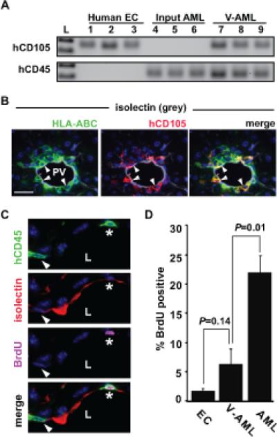
Figure 4. V-AML cells up regulate CD105 expression and become quiescent.

(A) Input primary AML cells express hCD45 but not hCD105 by RT-PCR. By contrast, V-AML cells sorted from the liver of an NSG recipient show an induction of CD105 expression. Each lane shows ten sorted cells. (B) hCD105 expression in a subpopulation of V-AML that are tightly associated with the endothelium. Arrowheads indicate hCD105+ (red), isolectin+ (grey) HLA-ABC+ (green) V-AML cells. DNA is labeled with DAPI (blue). (C) Example of BrdU uptake in PV-adherent V-AML cells. In the top panel, two hCD45+ (green) V-AML cells (*, arrowhead) that appear to share membrane with mouse endothelial cells are shown. The second panel shows mouse endothelial isolectin GS-IB4 expression (red). In the third panel, BrdU (pink) is detected in one of the two V-AML cells (*). Bottom panel is a merged image. L indicates the PV lumen. (D) Quantitation of BrdU uptake in mouse endothelial cells, AML and V-AML tightly associated to mouse CD31+ PV endothelium. VAML cells adherent to or incorporated into the endothelial layer of the PV proliferate significantly less than AML cells present throughout the rest of the liver. The mean ± SEM is shown for pooled samples. A minimum of 600 PV ECs, 75 PV integrated V-AML cells and 2500 AML cells were scored in non-adjacent sections from each liver. Scale bars: 20 microns
