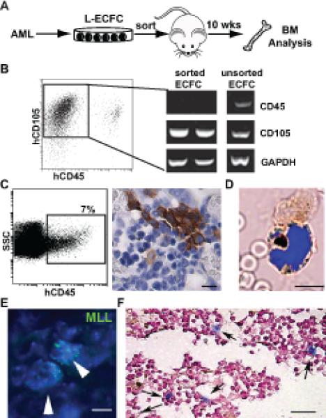
Figure 6. AML-derived endothelium can adopt a leukemic phenotype.

(A) Experimental strategy to assess in vivo leukemic potential of AML-derived ECFCs. (B) Flow cytometry gating used for the purification of CD105+CD45neg L-ECFC prior to transplant. RT-PCR analysis of sorted CD105+CD45neg cells confirmed the absence of CD45 expression. (C) Analysis of bone marrow from CD105+CD45neg L-ECFC recipient NSG mice. A significant population of human CD45+ cells is detected in mouse bone marrow by flow cytometry (left panel); and in tissue sections by immunohistochemistry (DAB brown). (D) Localization of hCD33+ expressing cells (brown) in the bone marrow of an L-ECFC engrafted NSG mouse. A representative merged brightfield and fluorescent image (DAPI, blue) is shown. (E) Multiple copies of human MLL gene (green, arrowheads) confirm the leukemic origin of the human donor cells in bone marrow from L-ECFC engrafted mice. All nuclei are stained with DAPI. (F) Detection of cytoplasmic NPM1 (blue, arrows) following transplant of L-ECFC derived from an NPM1 mutant donor patient. Scale bars: (C): 10 microns; (D-E): 5 microns; (F): 50 microns.
