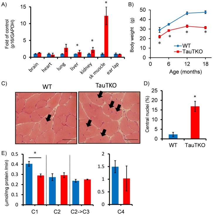
Figure 2. Accelerated aging in TauTKO muscle.
A) mRNA level of the senescence marker, p16INK4A, was quantified in several tissues of old TauTKO and WT mice by real-time RT-PCR. The marker is severely increased in old TauTKO muscle. n = 4–7. B) Body weight of TauTKO mouse is consistently lower than that of the WT mouse. n = 6–12. C), D) Histological analysis of tibial anterior muscles shows an induction of myotubes with central nuclei indicated by arrows in old TauTKO muscle. The ratio of myotubes with central nuclei to total myotubes is significantly higher in old TauTKO than WT. n = 6. Scale bar = 50 µm. E) The assays for electron transport chain complex reveal that activity of mitochondrial complex1 is decreased in old TauTKO muscle. C1–4; Complex 1–4. n = 4–5. *;p<0.05.

