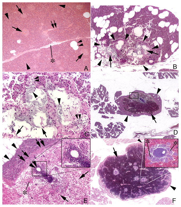
Figure 5.
Pancreatic tail histology. Panel A is from a control MetS pig and shows islets of Langerhans (arrows), pancreatic acinar cells (arrowhead), interlobular ducts (double arrow), fat cells (double arrowhead) and interlobular septum (asterisk). Panel B shows the first type of injury, which is focal (outlined by arrowheads) and characterized by cellular necrosis of acini (arrows). Panel C is at higher magnification and shows necrotic centroacinar cells (arrowheads), complete loss of acinar cells (arrows) and infiltrating inflammatory cells (double arrowheads). Panels D–F show the second type of injury, which is characterized by a focal intra-parenchymal hematoma (arrows) that is separated from surrounding normal appearing tissue (arrowheads) by a fibrotic band or capsule (double arrows in panel E which is an enlargement of the box shown in panel D). Islands of islets of Langerhans (asterisk), and various sized ductal and vascular profiles (double arrowheads) surrounded by chronic inflammatory cells are seen within the boundaries of the hematoma (Panels D–F). Inserts in panels E and F show chronic inflammatory cells around an artery. Insert in panel F also shows several islets of Langerhans (asterisk) near the vascular cuffs.

