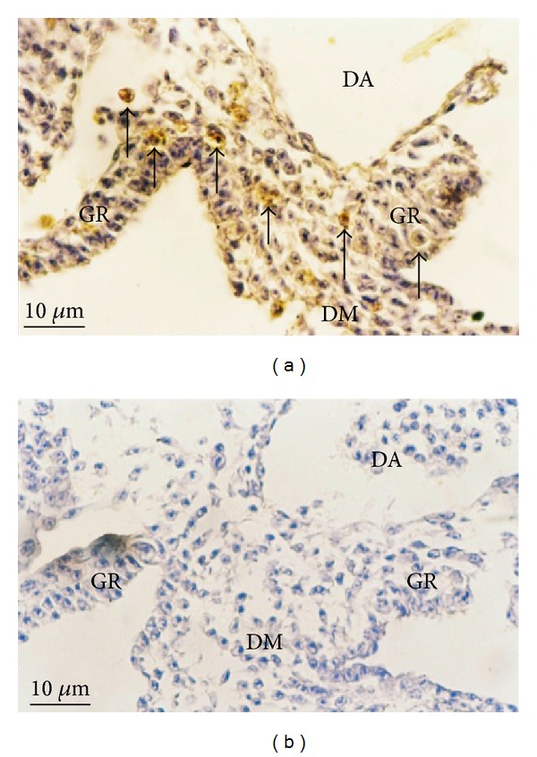
Figure 2.

Photomicrographs of 3-day-old quail embryos sectioned during genital ridge formation. (a) qPGCs and WFA positive cells (arrows) were detected in both left and right genital ridges (GR) as well as the dorsal mesentery (DM), but not in the dorsal aorta (DA) (lectin HC ×400); (b) no WFA positive cells in the negative control (×400).
