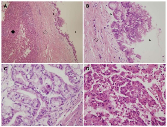
Figure 1.

Pathology of intrahepatic biliary cystadenoma and cystadenocarcinoma. A, B: Intrahepatic biliary cystadenoma (black diamond, hepatic tissue; hollow diamond, fibrous cyst wall; arrowhead, simple columnar epithelium; hollow arrowhead, cavity); C, D: Intrahepatic biliary cystadenocarcinoma. Mucinous cystadenocarcinoma with columnar epithelium, abundant cytoplasm, containing mucin, and nuclei located in the basal layer (C).
