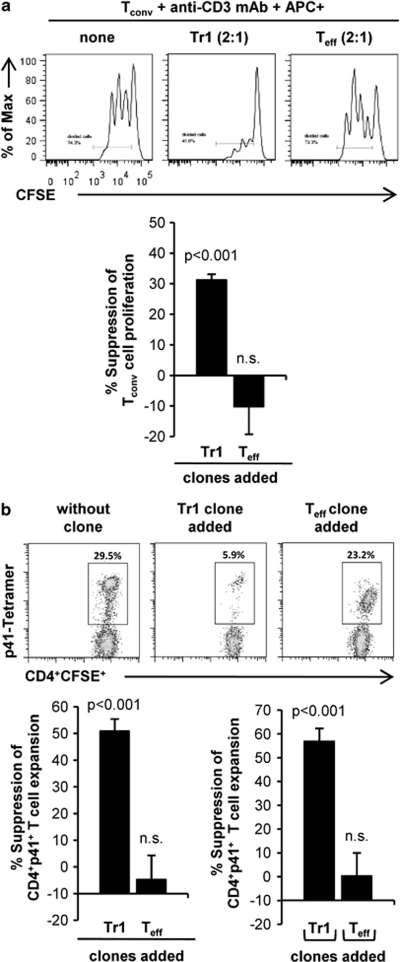
Figure 2.
Human p41+ Tr1 cell clones suppress expansion of CD4+ T cells. (a) T-cell clones were cocultured together with CFSE-labeled autologous CD4+CD25− T cells (Tconv) (1:2 ratio) and irradiated p41-pulsed CD2-depleted APC (1:2:4 (clone:Tconv:APC)) in round bottom plates, coated with 0.5 μg ml−1 anti-CD3 mAb. After three days, CFSE dilution was determined by flow cytometry. The histograms show CFSE dilution of Tconv in the absence and presence of one representative Teff and Tr1 clone. Diagrams summarize %±s.d. suppression of Tconv proliferation in the presence Tr1 and Teff clones (n=9 per group; three different donors) at a 1:2:4 ratio (clone:Tconv:APC). (b) CFSE-labeled PBMCs were cultured together with CellVue-Claret-labeled p41-specific T-cell clones at 2:1 ratio. After 24 h of culture and every second day, IL-2 were added to the culture. After seven days, the T cells were restimulated with p41-pulsed autologous DC at a ratio of 10:1 and cultured for additional six days in the presence of IL-7 and IL-15. Afterward, expansion of CFSE+CD4+p41+ T cells was analyzed by flow cytometry using a p41-specific PE-labeled tetramer. Dotplot analysis shows one experiment in the absence (left) and presence of a representative Tr1 clone (middle) and Teff clone (right). Diagrams summarize %±s.d. suppression of CD4+CFSE+p41+ T-cell expansion in contact-dependent presence of Tr1 and Teff clones (left) (n=6 per group; three different donors) as well as in contact-independent cocultures in transwell systems (right) (n=5 per group; three different donors).

