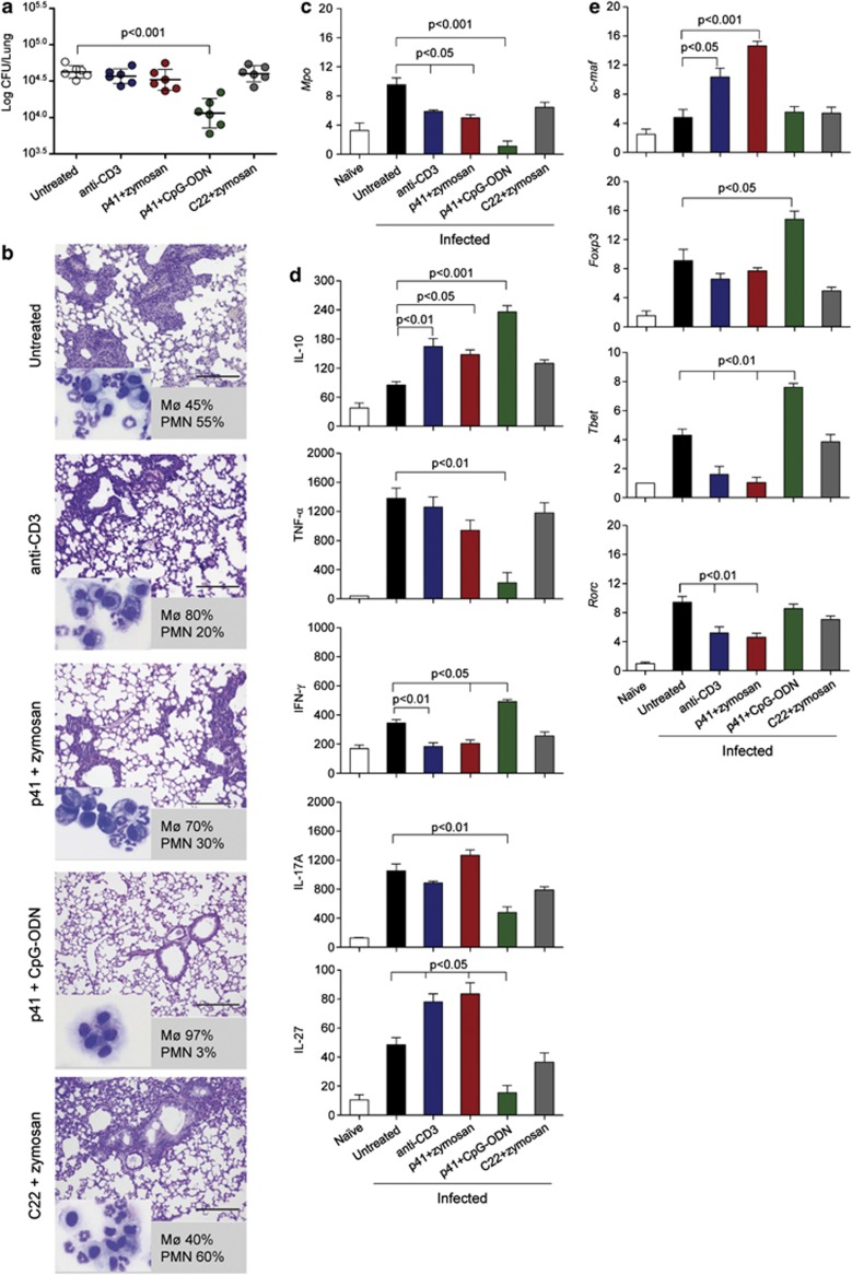
Figure 5.
p41-induced murine Tr1 cells decrease inflammatory T cells in murine aspergillosis. C57BL/6 mice were treated as decribed previously in Figure 3 and infected intranasally with live Aspergillus conidia (six mice per group). Mice were assessed for (a) lung fungal growth (log10 colony-forming unit per organ±s.d.), (b) lung histopathology (periodic acid-Schiff staining) and BAL cellular morphometry (indicated as % of mononuclear (Mø) or polymorphonuclear (PMN) cells in the inset (May–GrünwaldGiemsa staining)). Representative images of two independent experiments were acquired with a × 40 objective. Scale bars, 200 μm. (c) Mpo expression (reverse transcriptase-PCR) on total lung cells. (d) Levels of cytokines (pg ml−1) by specific enzyme-linked immunosorbent assays (mean values±s.d., n=3) in lung homogenates. (e) Relative expression (mRNA-fold increase) of transcription factor genes (by reverse transcriptase-PCR) on purified CD4+ T cells from TLN. Assays were done at three days post-infection. P-value was determined by one-way analysis of variance Bonferroni post-test (n=3).

