
Figure 3. Degradation of pGL3-PEI-coated nanoparticles.
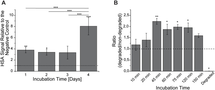
A: Dot blot immunoanalysis was used to evaluate the internalised HSA amount in Cb cells treated with pGL3-PEI-coated nanoparticles The HSA signal was related to the signal of the negative control (horizontal line). B: Similar conditions were used to evaluate the chemical degradation of pGL3-PEI-coated HSA nanoparticles with proteinase K. The HSA signal was related to the signal of non-degraded nanoparticles (horizontal line). Results are shown as mean ± S.E.M. (n = 3).