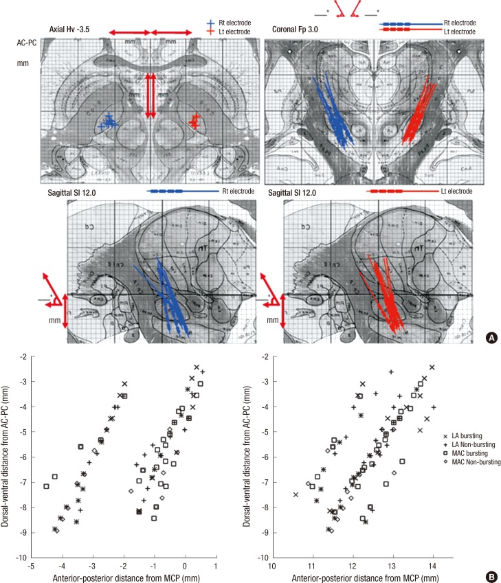
Fig. 4.
Location of the electrodes plotted onto the human brain atlas. Based on the CT-MRI fusion images of the preoperative brain MRI and postoperative brain CT scan taken one month after surgery. All the electrode positions are mostly located to the middle one third part of the STNs on both sides in the fused images. (A) It shows location of the electrodes plotted onto the human brain atlas of Schaltenbrand and Wahren. (B) The microelectrodes positions were plotted in the sagittal and coronal planes aligned along anterior commissure and posterior commissure line (AC-PC line).

