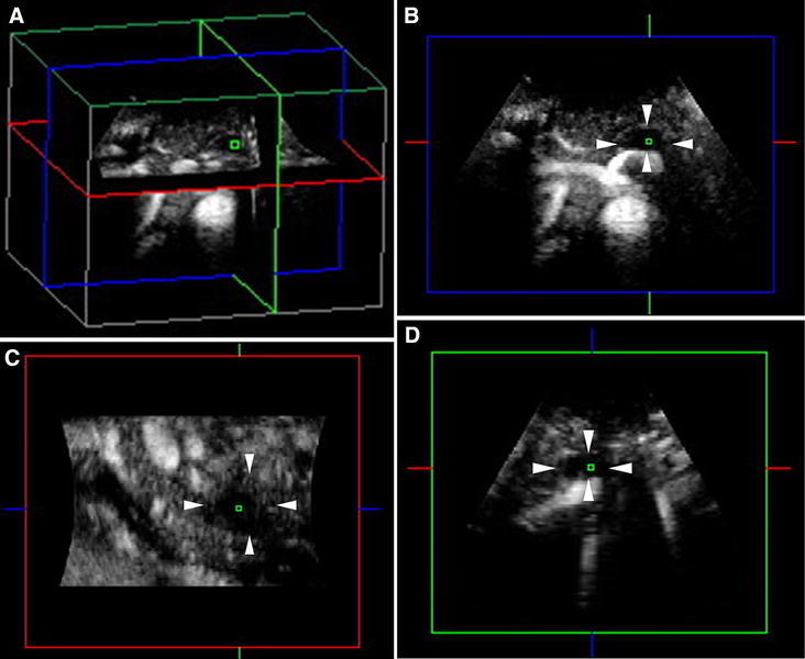
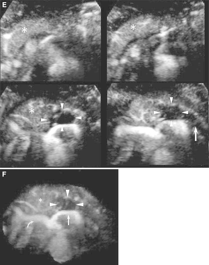
Fig. 2.


CE 3D US images acquired in the transverse plane of a 43-year-old man with PDAC (maximum diameter, 38 mm) in the pancreatic body. A–D Hypovascular lesions (arrowheads) are clearly seen in three orthogonal planes in the arterial phase. Small green squares show the center of a lesion. (A An image for navigation of the following planes; B plane A, from the front to the back; C plane B, from the left to the right; and D plane C, from the top to the bottom). E TUIs of CE 3D US (plane A) in the arterial phase show a hypovascular lesion (arrowheads) compared with the parenchyma in the pancreatic head (asterisks) with peritumoral and fine vessels. The dilated main pancreatic duct appears as a non-enhanced area in the caudal part of the lesion (arrow). F Sonographic angiogram in the arterial phase, rendered in the average intensity mode, shows the hypovascular lesion in the pancreatic body (arrowheads) compared with the parenchyma (asterisk). The splenic artery with encasement by tumor invasion (arrow) and common hepatic artery (curved arrow) is clearly seen posterior to the pancreas.
