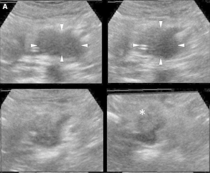
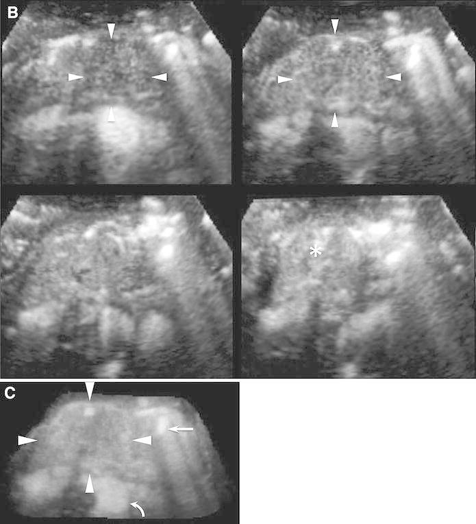
Fig. 3.


Conventional 3D US and CE 3D US images acquired in the transverse plane of a 78-year-old woman with MFP (maximum diameter, 33 mm) in the pancreatic head. A TUI of 3D US (plane A) in the B-mode shows a hypoechoic lesion in the pancreatic head (arrowheads). The image on the lower right shows the pancreatic parenchyma in the pancreatic body (asterisk). B TUIs of CE 3D US (plane A) in the arterial phase show an isovascular lesion (arrowheads) compared with the pancreatic parenchyma (asterisk). Dense fine vessels are seen in a whole lesion, hypoechoic on B-mode US, similar to the pancreatic parenchyma. C Sonographic angiogram in the arterial phase, rendered in the average intensity mode, shows homogeneous enhancement (arrowheads) of both the pancreatic head and body. Superior mesenteric artery (arrow) and abdominal aorta (curved arrow) appear around pancreas.
