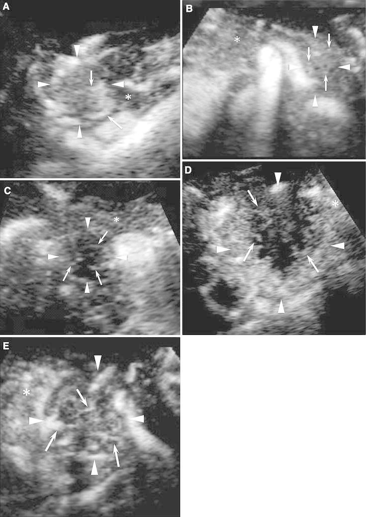
Fig. 4.

CE 3D US images showing the typical enhancement patterns of solid pancreatic lesions. A A hypervascular lesion (arrowheads) compared with the pancreatic parenchyma (asterisk) with intratumoral and irregular vessels (arrows). B An isovascular lesion (arrowheads) compared with the pancreatic parenchyma (asterisk) with intratumoral and fine vessels (arrows). C A hypovascular lesion (arrowheads) compared with the pancreatic parenchyma (asterisk) with peritumoral and fine vessels (arrows). D A hypovascular lesion (arrowheads) compared with the pancreatic parenchyma (asterisk) with peritumoral and irregular vessels (arrows). E Hypovascular lesions (arrowheads) compared with the pancreatic parenchyma (asterisk) with intratumoral and irregular vessels (arrows).
