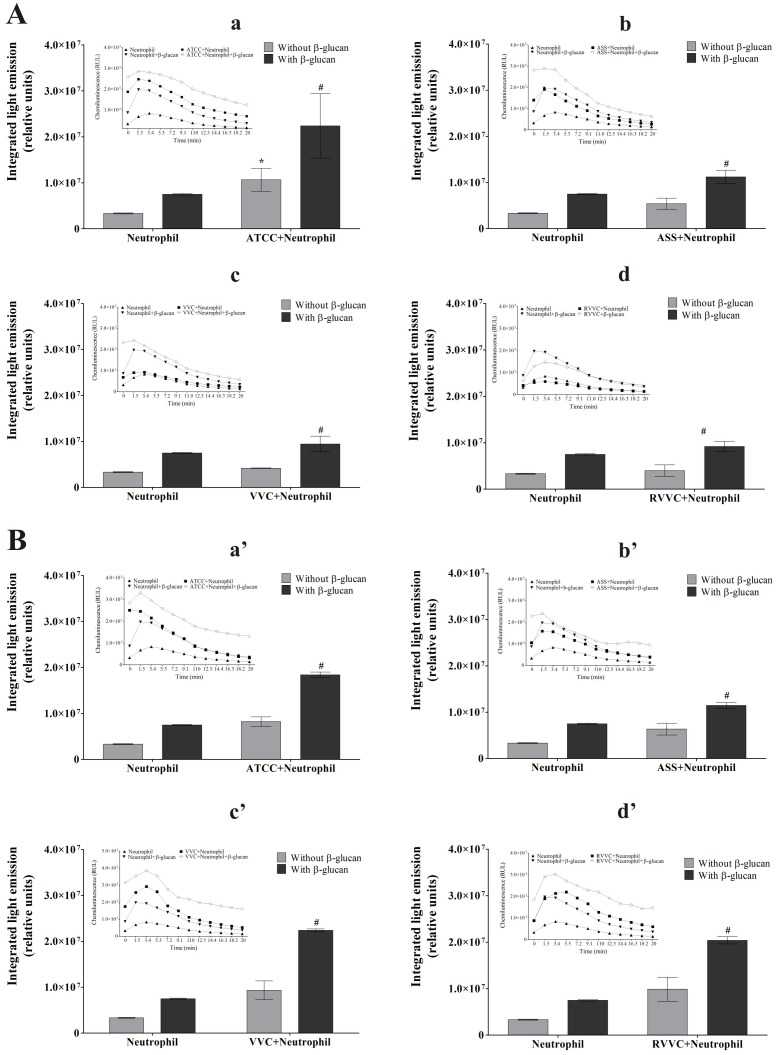
Figure 5. Myeloperoxidase activity of β-glucan-treated neutrophils activated by different isolates of C. albicans and C. glabrata (integrated light emission).
The inset represents kinetic study of MPO activity of β-glucan-treated neutrophils after 20 minutes of incubation. Neutrophils (2.0×10⁶ cells/ml) were previously treated or not with 3 mg/ml β-glucan and incubated with the reference strain and different isolates of (A) C. albicans and (B) C. glabrata (2×10⁷ CFU/ml) for 30 min. (a,a’) ATCC. (b,b’) ASS. (c,c’) VVC. (d,d’) RVVC. After incubation, chemiluminescence was monitored for 20 min at 37°C in a microplate luminometer using luminol as a chemical light amplifier. The data are expressed as the mean ± SD of three independent experiments. *p≤0.05, significant difference compared with the control group (neutrophils alone); # p≤0.05, significant difference compared with untreated and activated neutrophils.

