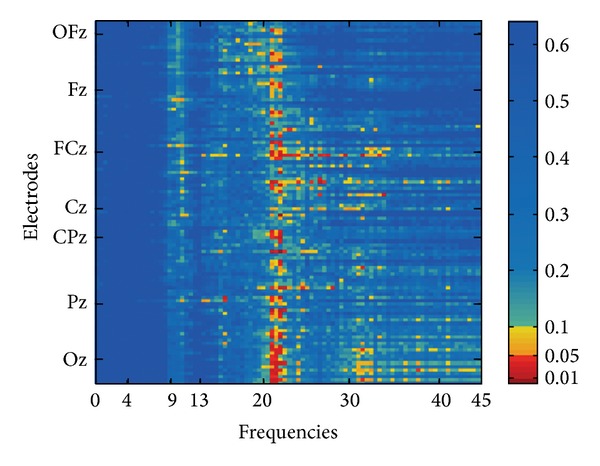
Figure 2.

Comparison of TI versus CO normalized EEG spectral power adjacency matrix of the group comparison (TI versus CO): significance levels (P values) of the correlations are color-coded (one-sided). On the y-axis electrodes are aligned from rostral (top) to caudal (bottom), irrespective of laterality. Positions of electrodes on the central line are marked.
