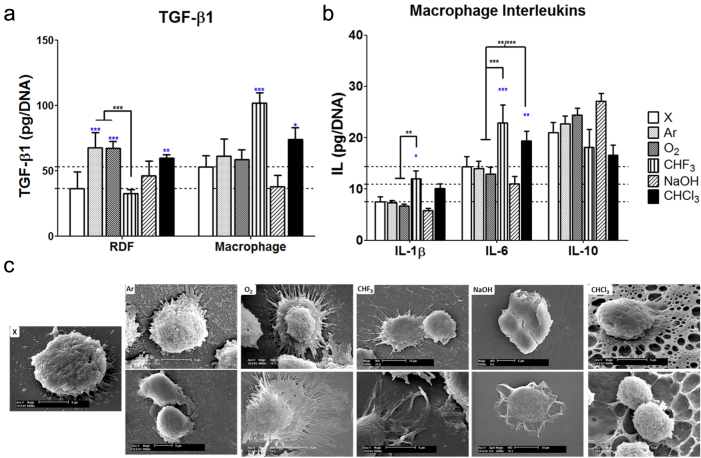
Figure 4. Cytokine analysis on conditioned medium co-culture in vitro studies.
(a) TGF-β1 secretion in RDFs and macrophages at day 1. (b) Interleukin (IL-1β, IL-6 and IL-10) expression in macrophages in mono (ref) or conditioned medium co-cultures (CM) at day 1. Normalized per DNA represent amount secreted for 100,000 cells. Each horizontal tick is in reference to TGF-β1 RDFs and macrophages (a), and IL-1β, IL-6 and IL-10 macrophage (b) value of unmodified rods. Data are shown as mean ± s.d. (n = 3). Blue stars (*P < 0.05, ** P < 0.01, *** P < 0.001) indicate statistical significances in comparison to unmodified, and black stars evaluate statistical differences between the different treatments. (c) SEM images (scale bar: 5 and 10 μm) show different macrophage morphology on different surface treatments in mono-culture study. Filopodia of macrophages seeded on nano-scale modification of Ar, O2, and CHF3 were more extended compared to CHCl3 providing micro-scale modification on its surface, and NaOH providing no surface topography modification.

