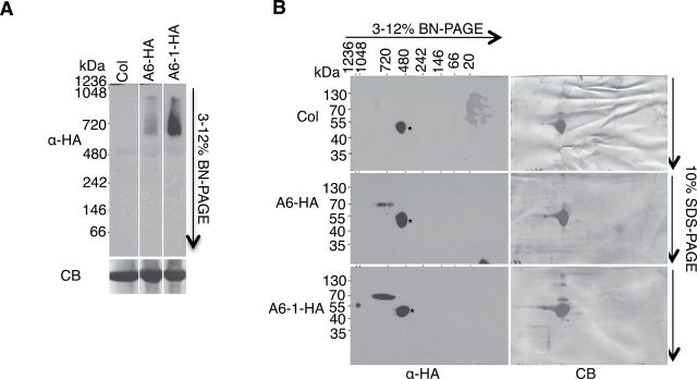
Figure 2.

ACD6-HA and ACD6-1-HA Reside In Large Complexes.
Microsomal proteins from the indicated genotypes were solubilized with 0.5% Triton X-100 and separated in the first dimension by BN–PAGE (A) and in the second dimension by SDS–PAGE (B), and analyzed by immunoblotting with HA antibody. CB, Coomassie blue-stained membrane; *, non-specific band. Lanes in (A) were from one continuous membrane. These experiments were repeated three times, with similar results.
