Figure 1.
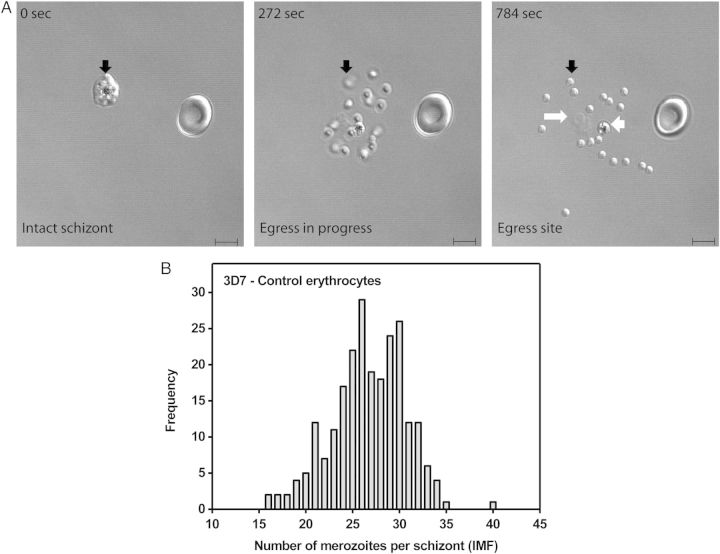
P. falciparum 3D7 schizonts produce variable numbers of merozoites in control erythrocytes. A, Formation of a merozoite egress site by a 3D7 schizont infected control erythrocyte. Selected frames from a time-lapse recording of the egress process are shown. The 0-second frame shows an uninfected erythrocyte and an intact segmenting schizont (black arrow) containing multiple merozoites, which cannot be accurately counted. The 272-second frame shows merozoites in motion shortly after egress. Those that have not yet settled onto the glass surface are out of focus (black arrow). The 784-second frame shows a fully formed egress site littered with a single food vacuole (short white arrow) containing hemozoin, erythrocyte membrane fragments (long white arrow), and 18 merozoites (short black arrow), which have scattered and settled onto the glass surface. The “intraerythrocytic multiplication factor” (IMF) of this particular schizont is 18. DIC microscopy, scale bar = 5 µm. B, Non-Gaussian frequency distribution of 3D7 IMFs for 237 singly infected control erythrocytes. Data are derived from 17 independent experiments using erythrocytes from 13 healthy donors.

