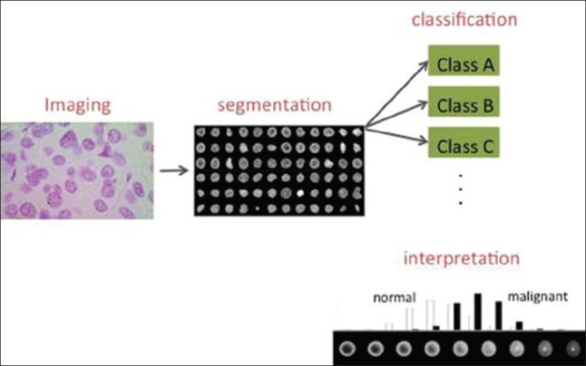
Figure 1.

Nuclear structure extraction and quantification process. A Feulgen stained tissue section from a patient suspected of having fetal-type hepatoblastoma. Nuclei are first automatically segmented, and then utilized for cancer detection and subtyping using classification approaches. Modern mathematical algorithms for image analysis are also able to display intuitive visualizations depicting differences between nuclear classes. In this case, malignant cell distributions, on average, tend to have their chromatin more evenly distributed throughout the nuclear envelope
