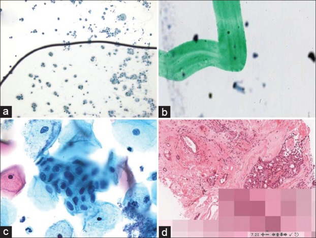
Figure 3.

Digital pathology image aberrations. (a) An air bubble on this slide has caused many of the cells to be out of focus. (b) The green dotting pen mark on this slide is in focus whereas the cells are not. (c) Not all of the endocervical cells in this cell group are in focus because this Pap test slide was scanned with a single z plane. (d) Pixilated image due to slow internet connectivity
