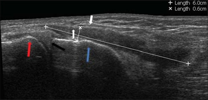
Figure 7.

39-year-old man who suffered from pain in the medial aspect of the distal right knee diagnosed with Pes anserine bursitis. Extended panoramic ultrasound view of the medial part of the knee shows fluid in the pes anserinus bursa (asterisks), that was located between the pes anserine ligament (white arrows), medial condyle of the femur (red arrow), medial meniscus (black arrow), and medial distal part of the tibia (blue arrow).
