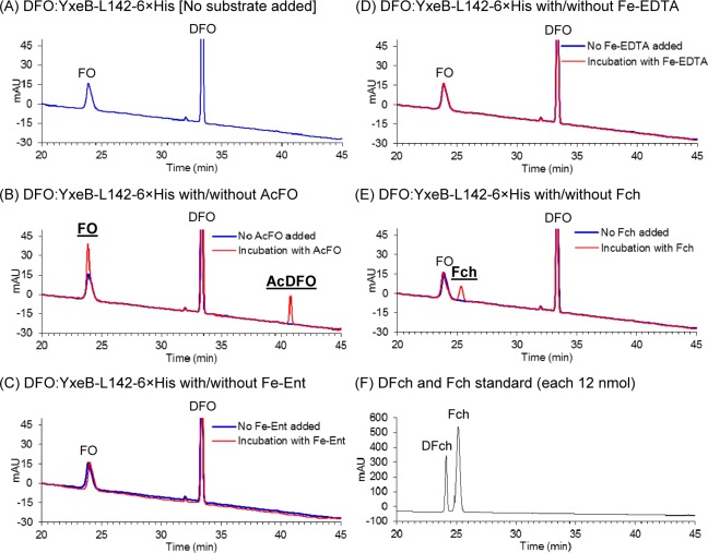
Figure 5.
Iron exchange from Fe-siderophores or iron-chelators to the DFO:YxeB-L142-6×His complex. (A to E) After 4 μM DFO and 2 μM the YxeB protein had been mixed to create the DFO:YxeB complex, 0.2 μM AcFO (panel B), Fe-Ent (panel C), Fe-EDTA (panel D), or Fch (panel E) was added to the sample. The complex without substrate addition (blue chromatograph) and the complex after 40 min incubation with the substrate (red chromatograph) were collected and analyzed by RP-HPLC. Peaks with the thick and underlined letters are the increased products. The calculated amount of compounds bound to the protein is shown in Table 2. (F) RP-HPLC analysis of DFch and Fch standards.

