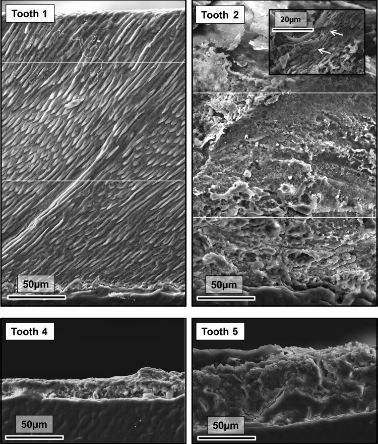
Figure 4.
SEM of representative exfoliated teeth. Tooth 1 exhibits normal enamel architecture comprising prisms (rods) of individual enamel crystallites classically described in numerous research publications and text books. The SEM representative of the thicker enamel found covering tooth 2 is lacking the obvious long range order and structure that characterizes normal enamel. However, very occasional areas exhibiting prism-like structures are present (indicated by arrows on inset micrograph). Where present, the enamel covering teeth 4 and 5 is thin and exhibits no structural similarities to normal enamel. The white horizontal lines show where separate micrographs have been collaged to create a wider field of view.

