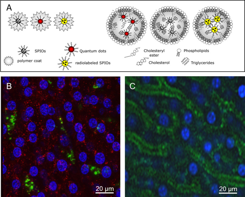
Figure 1.
Characterization of nanocrystals and uptake into liver cells in vivo. (A) Oleic acid-stabilized SPIOs, radiolabeled SPIOs (59Fe-SPIOs) or QDs are embedded in PMAOD-polymer or in lipid micelles as indicated in the model. (B) Native DID-labeled LDL (red) and QDs-micelles (green) or (C) polymer-embedded QDs (green) were injected into wild type C57BL/6 mice via a tail vein catheter. Nuclei were stained by intraperitoneal injection of the fluorescence dye Hoechst 33342. 30 min after injection, the liver was excised and directly placed on the confocal microscope. As shown in (B), LDL are internalized by hepatocytes (red) whereas signals from QDs embedded within lipid micelles are found associated with hepatic Kupffer cells (green). (C) shows that polymer-embedded QDs are primarily associated with endothelial cells lining the sinusoids of the liver. Scale bars = 20 µm.

