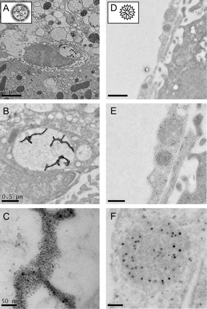
Figure 2.
Cryo-electron microscopy of hepatic nanocrystals uptake. SPIOs-micelles (left panel) or polymer-embedded SPIOs (right panel) were injected into wild type C57BL/6 mice. 30 min after injection, mice were perfused with 2% paraformaldehyde in PBS and livers were processed for electron microscopy. (A–C) The pictures highlight a Kupffer cell 30 min after the injection of SPIOs-labelled lipid micelles. Clustered nanocrystals of lipid micelles can be detected within intracellular compartments, probably a lipid droplet-like structure of the cell. (D–F) Polymer-coated SPIOs can be found in endosomal structures of endothelial cells. Scale bars correspond to 2 µm (A,D), 0.5 µm (B,E) and 50 nm (C,F), respectively.

