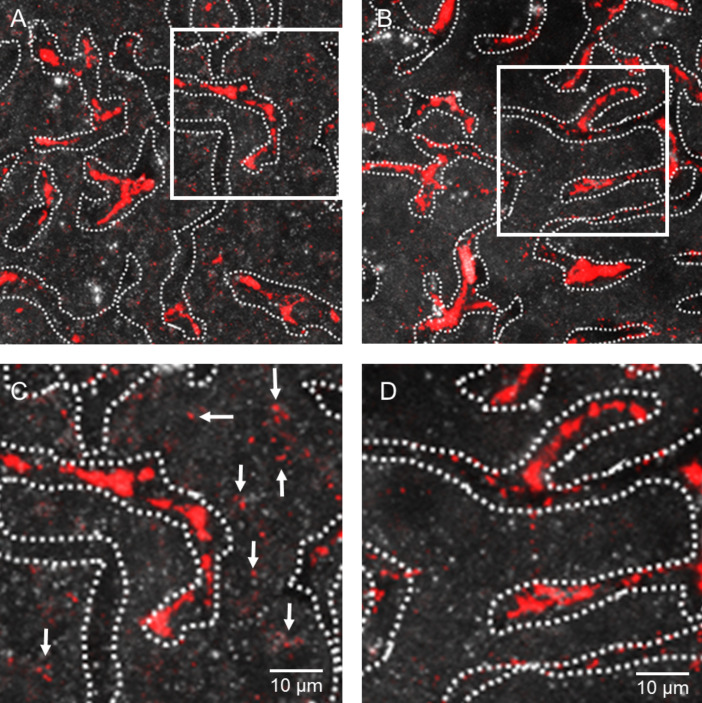
Figure 5.
QDs-micelles uptake into hepatocytes is dependent on apolipoprotein E. QDs-micelles (red) were injected into wild type mice (left panel) or into apolipoprotein E-deficient mice (right panel). 30 min after injection, livers were excised and directly analysed by using confocal imaging. Liver sinusoids were visualized by the reflection mode in the unstained tissue and the capillary lumen is surrounded by dashed lines. In the wild type situation, QDs-derived fluorescence were found in Kupffer cells, which are located within the lumen of liver sinusoids (A). In addition, high magnification of the highlighted square revealed uptake of QDs-micelles into hepatocytes (C, indicated by the arrows). In apolipoprotein E-deficient mice, QDs can mainly be detected within the lumen of liver sinusoids (B,D), which indicates that the QDs-micelle uptake into hepatocytes is dependent on apolipoprotein E.

