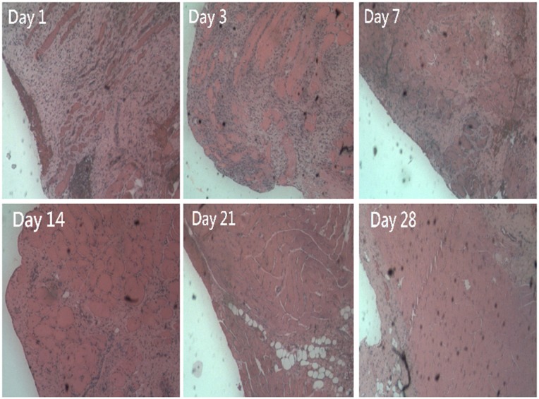
Figure 9.
Histological examination of muscle tissue surrounding the plate at days 1, 3, 7, 14, 21, and 28 after surgery. Microscopic examination of hematoxylin-and-eosin-stained specimens showed significant mononuclear cell infiltrates of lymphocytes, plasma cells, and eosinophils in the muscle tissue surrounding the plate at day 1 after surgery. The number of polymorphonuclear leukocytes gradually decreased over time to day 28 post-surgery.
Note: Original magnification 200×.

