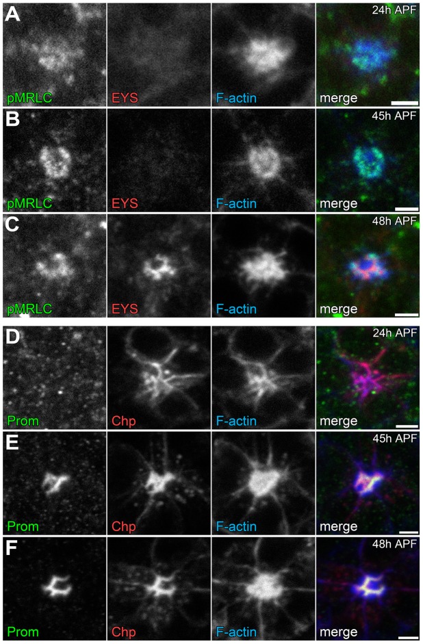
Figure 8. Temporal profile of the coordination of actomyosin contraction, steric hindrance of adhesion, and secretion of an extracellular matrix.
(A–F) Confocal immunofluorescence micrographs of wild-type ommatidium. (A–C) Ommatidium stained with phospho-Sqh (pMRLC, green), EYS (red), and F-Actin (blue) at: (A) 24 h APF. (B) 45 h APF. (C) 48 h APF. (D–F) Ommatidium stained with Prominin (Prom, green), Chaoptin (Chp, red), and F-Actin (blue) at: (D) 24 h APF. (E) 45 h APF. (F) 48 h APF. Scale bar, 2 µm.

