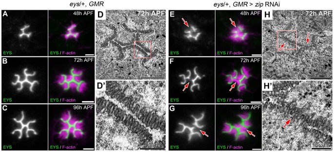
Figure 9. Defects in apical membrane separation are detected early and maintained throughout development.
(A–C, E–G) Confocal immunofluorescence micrographs of (A–C) eys, GMR-Gal4/+ ommatidium and (E–G) eys, GMR-Gal4/+; UAS-zip RNAi/+ ommatidium co-stained with EYS (green) and F-actin (magenta) at (A,E) 48 h APF. (B,F) 72 h APF. (C,G) 96 h APF. (D,H) Transmission electron micrograph of (D,D′) eys, GMR-Gal4/+ ommatidium and (H,H′) eys, GMR-Gal4/+; UAS-zip RNAi/+ ommatidium at 72 h APF, (D′,H′) represents high magnification of boxed region in (D) or (H), respectively. Arrows denote juxtaposed apical rhabdomere membranes. Scale bar, (A–D, E–H) 2 µm, (D′,H′) 0.5 µm.

