
Figure 3. Neointimal cells in AVF are originated from cells in the vein.
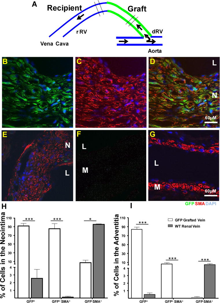
A. Diagrammatic representation of the composite AVF created by interposing a GFP vena cava between the recipient renal vein and the aorta of a WT recipient. The area used for cross sections appears between discontinuous lines.
B–D. Representative pictures of neointimal cells in the grafted vein. Most of those cells stained positively for GFP and SMA which revealed that those cells were originated from local cells in the graft. Microphotographies from the GFP (green, B) and SMA (red, C) channels were overlaid on Figure D.
E. Representative section from the recipient renal vein. This section was taken from an area close to the vein-to-vein anastomotic point. Most of the neointimal cells were SMA+ (red) GFP−(green). Only rare GFP+ cells from the graft were found infiltrated in this part of the neointima.
F and G. Sections from the recipient aorta and the vena cava showed the absence of tissue autofluorescence and neointima in those vessels. Vascular smooth muscle cells were stained with an antibody against SMA (red). No green autofluorescence was detected. Nuclei were counter stained with DAPI (blue). M: Media; N: Neointima; L: Lumen.
H and I Histograms showing the number of GFP and GFP SMA positive and negative cells in the neointima and adventitia of the composite fistula. Bars represent the mean ± SEM, n=3. ***p<0.01; * p<0.05.