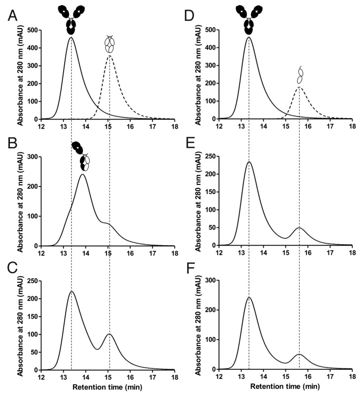
Figure 5. HPLC traces analyzing Fab arm exchange. (A) Overlay of separate runs for a wild-type IgG4, represented by the black cartoon image, and an IgG4 wild-type hinged Fc domain, represented by a white cartoon image. (B) A mixture of IgG4 antibody and IgG4 wild-type hinged Fc after incubation at 37°C for 96 h in the presence of 0.5 mM GSH. The wild-type Fc undergoes arm exchange with the IgG4 antibody to produce an intermediate sized heterodimer, as indicated by the cartoon representation. (C) The same respective incubation as (B) but in the absence of GSH showing no arm exchange. (D) Overlay of separate runs for the IgG4 antibody and a monomeric hinged Fc domain with the T394D mutation. (E) A mixture of IgG4 antibody and monomeric Fc after incubation at 37°C for 96 h in the presence of 0.5 mM GSH. No arm exchange is detectable. (F) The same respective incubation as (E) but in the absence of GSH.

An official website of the United States government
Here's how you know
Official websites use .gov
A
.gov website belongs to an official
government organization in the United States.
Secure .gov websites use HTTPS
A lock (
) or https:// means you've safely
connected to the .gov website. Share sensitive
information only on official, secure websites.
