
Figure 2. Pho23 Negatively Regulates Autophagy Activity.
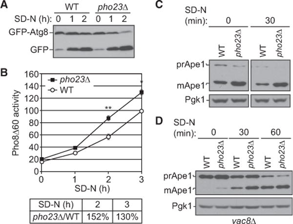
(A) Wild-type (BY4742) and pho23Δ (JMY018) cells with a centromeric plasmid expressing CUP1 promoter-driven GFP-ATG8 were grown to mid-log phase inSMD-Ura and shifted to SD-N for the indicated times. Autophagy activity was measured by the GFP-Atg8 processing assay.
(B) Wild-type (WLY176) and pho23Δ (JMY048) cells were grown to mid-log phase in YPD and shifted to SD-N for the indicated times of nitrogen starvation, and autophagy activity was monitored by the Pho8Δ60 assay. Pho8Δ60 activity was normalized to the wild-type strain (set to 100%) after 3 hr of nitrogen starvation. The graph shows the average activity from three different experiments. Error bars represent the SEM. Two-tailed paired t test was used for statistical significance; *p < 0.05, **p < 0.01.
(C) Wild-type (SEY6210) and pho23Δ (JMY047) cells were grown overnight, diluted to 0.1 optical density 600 (OD600), grown to mid-log phase (0.6 OD600) in YPD, and shifted to SD-N for 30 min of nitrogen starvation. The precursor (pr) and mature (m) forms of Ape1 were separated by SDS-PAGE and detected with anti-Ape1 antiserum by western blotting. Pgk1 was detected with anti-Pgk1 antiserum as a loading control.
(D) Precursor Ape1 processing in wild-type (vac8Δ; CWY230) and pho23Δ (pho23Δ vac8Δ; JMY146) cells at 0, 30, and 60 min after nitrogen starvation was detected by western blotting as in (C).