
Figure 7.
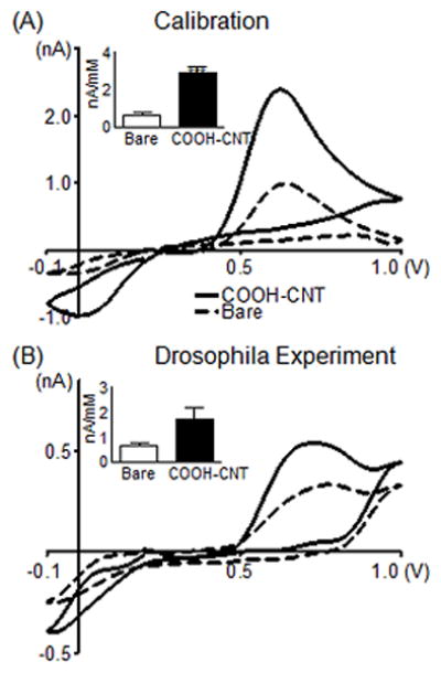
Measurements of endogenous serotonin in Drosophila. (A) Effect of COOH-CNT modification on the detection of 10 μM serotonin using the “serotonin waveform.” Electrode was scanned from 0.2 to 1.0 to −0.1 and back to 0.2 V at 1000 V/s at 10 Hz. The CV compares an electrode before (dashed) and after (solid) CNT modification during in vitro calibration. The inset shows average sensitivity for the oxidation peak (n=5). (B) Detection of serotonin in a larval Drosophila melanogaster ventral nerve cord. Different nerve cords are used for the bare (dashed) and COOH-CNT (solid) electrodes. Converting current to concentration using calibration data shows the COOH-CNT detected 650 nM serotonin with a peak oxidation current of 0.54 nA, while the bare electrode detected 870 nM serotonin with a peak oxidation current of 0.33 nA. The inset shows the average sensitivity for bare electrodes and COOH-CNT modified electrodes in Drosophila (n=5).