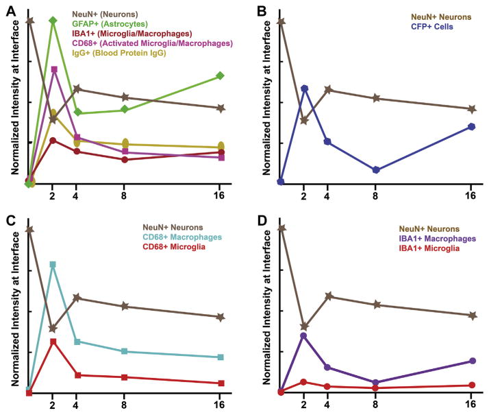
Fig. 11.
Summary of immunohistochemical assessment following microelectrode implantation. (A) Time course of neuroinflammatory markers and neurodegeneration following microelectrode implantation demonstrates no correlation between IBA1+, CD68+ or IgG + immunoreactivity and neuronal dieback. However, GFAP+ immunoreactivity was highest at two and sixteen weeks, the same time points we observed neurodegeneration. (B) We observed a temporal correlation between increased CFP+ immunoreactivity and neurodegeneration following microelectrode implantation. (C) In evaluating co-localization of activated macrophages and activated microglia within the CD68+ population, we did not observe a correlation between either cell type (activated microglia or activated macrophages) and neurodegeneration. (D) In evaluating the co-localized population of macrophage versus microglia within the total IBA1+ population, we observed a correlation between macrophages, but not microglia and neuronal dieback.

