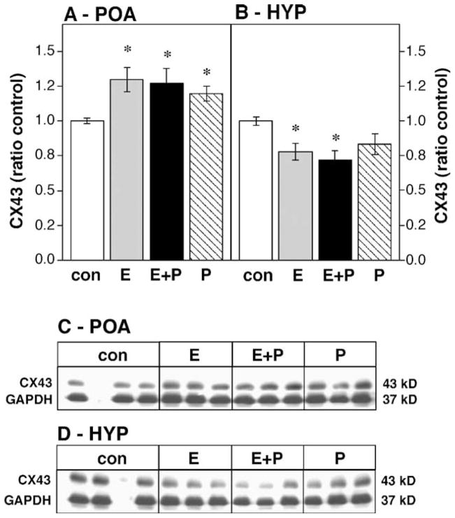
Fig. 1.
Effects of steroid hormones on CX43 protein levels in the OVX female POA (A) and HYP (B) expressed as a ratio to oil-injected controls (con, white bars). Hormone treatments include estradiol benzoate for 48 h (E, gray bars), progesterone for 2 or 4 h (P, hatched bars) or the combination of estradiol for 48 h followed by progesterone for 2 or 4 h (E + P, black bars); n = 15 in each group. * Significantly different from control ( P < 0.05). Data are expressed as means ± standard error for this and the following graphs. (C and D) Representative Western blots showing CX43 and GAPDH bands for the female POA and HYP, respectively. The blank lane in the control condition contained molecular weight markers.

