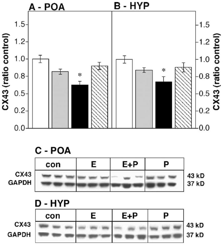
Fig. 2.
Effects of steroid hormones on CX43 protein levels in the castrated male POA (A) or HYP (B) expressed as a ratio to oil-injected controls; n = 4–5 in each group. For abbreviations, see Fig. 1. * Significantly different from control (P < 0.05). (C and D) Representative Western blots showing CX43 and GAPDH bands for the male POA and HYP, respectively.

