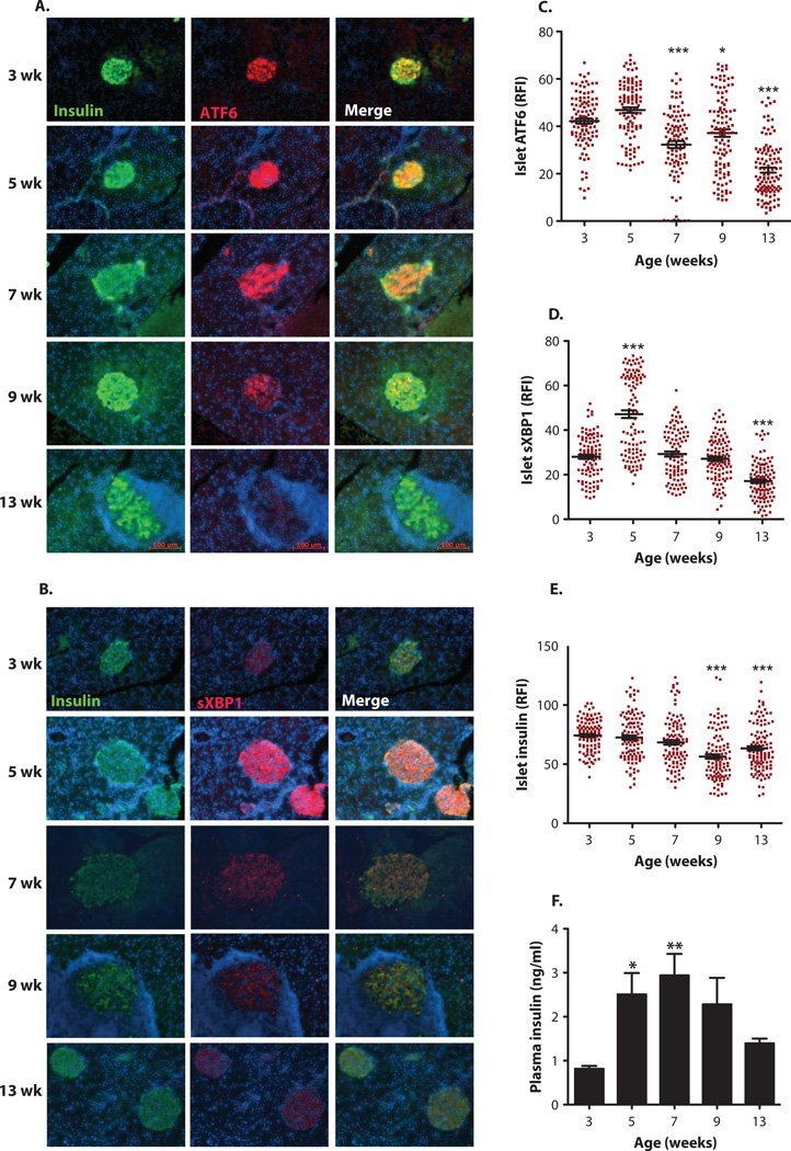
Fig. 1. Time-course detection of altered expression of UPR mediators in the islets of NOD mice.
Female NOD mice (n = 14 for each group) were sacrificed at 3,5,7,9, and 13 weeks of age. (A and B) Immunofluorescence assays were performed on the pancreatic sections by costaining with either (A) anti-ATF6 antibody (red) or (B) anfrsXBPI (red) and anti-insulin (green) antibodies. The cell nuclei were counterstained with 4’,6-diamidino-2-phenylindole (DAPI) (blue). (C to E) Relative fluorescence intensity (RFI) for (C) ATF6, (D) sXBPI, and (E) insulin was calculated by MATLAB (15 to 25 islets per animal per time point). (F) Serum insulin levels of NOD mice (n = 14) were measured by enzyme-linked immunosorbent assay (EUSA). All data are presented as means ± SEM, with statistical analysis performed by one-way analysis of variance (ANOVA) (***P < 0.001; **P < 0.005; *P < 0.05).

