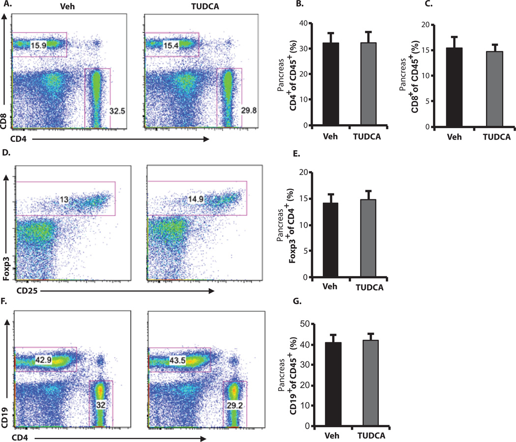
Fig. 4. Lack of TUDCA-induced alterations in immune cell populations in NOD pancreata.
Ten-week-old female NOD mice were treated with TUDCA (500 mg/kg per day) or vehicle (PBS) (n = 7 in each group) for 4 weeks. Each pancreas was isolated and dispersed, and cells were analyzed by flow cytometry after staining with antibodies against CD45, CD4, CD8, CD19, CD25, and Foxp3. Cells were pregated as CD45+. (A) Fractions of CD4+ or CD8+ T cells in representative dot plots. (B and C) Summary data for all mice. (D and E) Corresponding dot plots and summary data for Foxp3+ cells pregated as CD45+ and CD4+. (F and G) Corresponding dot plots and summary data for CD19+ B cells pregated as CD45+. Student′s t test did not show any significant statistical difference between vehicle- and TUDCA-treated cells.

