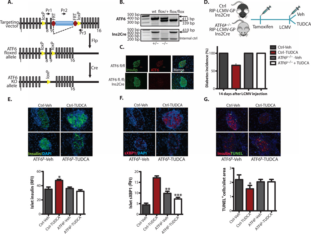
Fig. 7. TUDCA protection of β cells through the ATF6 branch of the UPR.
(A) Schematic representation of a targeting vector used for the generation of conditional β cell-specific ATF6-deficient (ATF6β−/−) mice. (B) Genotyping of control and ATF6β−/− mice was performed with tail DNA. (C) An immunofluorescence assay was performed to validate the tissue-specific deletion of ATF6. (D) Vehicle (PBS) or TUDCA (250 mg/kg twice daily via intraperitoneal injection) was applied to ATF6 wild-type (wt) control mice or ATF6β−/− RIP-LCMV-GP mice immediately after induction of diabetes with LCMV (2 × 10³ PFU/ml), and blood glucose levels were monitored for 2 weeks. (E) Female control and ATF6β−/−; RIP-LCMV-GP; lns2Cre mice were treated with either vehicle or TUDCA (n = 4 each group) for 2 weeks. (F) The mice were then sacrificed, and the relative immunofluorescence intensity (RFI) of insulin (green) or sXBPI (red) expression (20 to 30 islets per animal per group) was quantified by MATLAB. (G) Pancreatic sections from vehicle- or TUDCA-treated control and ATF6β−/− RIP-LCMV-GP mice (n = 4 each group, 20 to 30 islets per group) were examined for apoptosis with the TUNEL assay. All data are presented as means ± SEM, with statistical analysis performed by one-way ANOVA (***P < 0.001; **P < 0.005; *P < 0.05).

