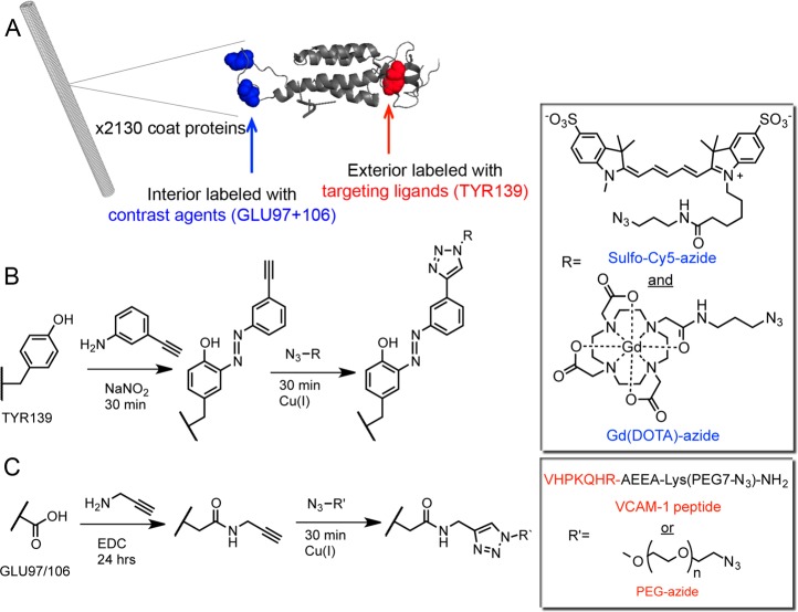
Figure 1.
(A) An illustrative image (PyMol and Chimera) of the structure of tobacco mosaic virus rods and its coat protein. The exterior (red) and interior (blue) reactive amino acids are highlighted in the individual coat protein. Bioconjugation of VCAM-1 targeting ligands or PEG and contrast agents to the surface of TMV involved the following sequence of reactions: (B) exterior incorporation of alkynes followed by attachment of VCAM-1 or PEG, followed by (C) internal channel incorporation of alkynes and contrast agent modification. Chemical structures are shown in the boxes.

