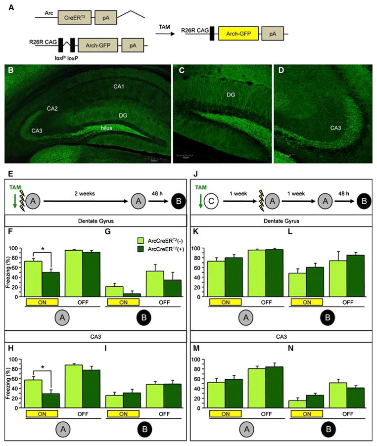
Figure 5. In Vivo Optogenetic Inhibition of the DG and CA3 Impairs Expression of Initially Encoded Memory.
(A) Genetic design.
(B–D) Representative images of the (B) hippocampus (the scale bar represents 200 μm), (C) DG (the scale bar represents 100 μm), and (D) CA3 (the scale bar represents 100 μm).
(E) The experimental design consisted of all mice being trained in four-shock CFC paradigm and then being re-exposed to context A and B Each context exposure consisted of 3 min of laser stimulation and 3 min without laser stimulation.
(F and G) Optogenetic inhibition of Arch-GFP+ DG neurons impaired expression of the corresponding fear memory in context A (F) in ArcCreERT2(+) mice when compared with ArcCreERT2(−) mice (p = 0.02) but had no effect in (G) context B (n = 6–9 mice per group).
(H and I) Optogenetic inhibition of Arch-GFP+ CA3 neurons impaired expression of the corresponding fear memory in context A (H) in ArcCreERT2(+) mice when compared with ArcCreERT2(−) mice (p < 0.02) but had no effect in (I) context B (n = 9–12 mice per group).
(J–L) Experimental design. Optogenetic inhibition of Arch-GFP+ DG neurons labeled in context C (J) did not impair freezing in ArcCreERT2(+) mice when compared with ArcCreERT2(−) mice in (K) context A or in (L) context B (n = 4–5 mice per group).
(M and N) Optogenetic inhibition of Arch-GFP+ CA3 neurons labeled in context C had no effect on memory expression in ArcCreERT2(+) mice when compared with ArcCreERT2(−) mice in (M) context A or in (N) context B (n = 5 mice per group). Error bars represent ± SEM.
See also Figures S4 and S5.

