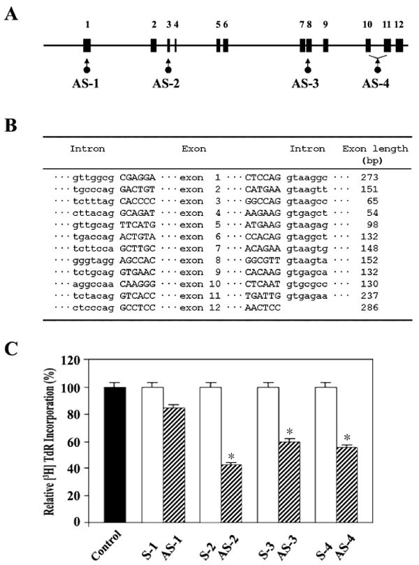
Fig. 5.

Exon-intron boundary (A) and genomic organization (B) of murine PHGDH gene, and Western blot analysis of the lysate of G0 T cells activated for 40 h by immobilized anti-CD3 with either no treatment or after treatment with PHGDH antisense or sense oligonucleotides. Murine G0 T cells were pretreated with individual antisense or sense phosphorothioate oligonucleotides at 25 μM for 4 hr before activation by immobilized anti-CD3 for 40 h. For [3H]TdR incorporation assay, the last 2 h was pulsed with 1 μCi of [3H]TdR before the cells were harvested. Each value is expressed as mean ± SD (n = 3). *P < 0.05 as compared with the control.
