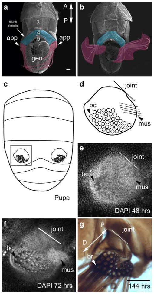
Fig. 1.
The abdominal appendages of T. biloba are located on the fourth segment of the male abdomen. a An SEM of the ventral side of the adult male abdomen shows the paired abdominal appendages (false-colored fuschia) are attached to the lateral edge fourth sternite (false-colored blue). b These abdominal appendages have a joint and musculature, which allows for 180° rotation. c The abdominal appendages first appear during pupation. The gray box indicates the abdominal appendage shown in d–g. d A cartoon of the abdominal appendage illustrates its morphological features: a large field of bristle cells (bc), a joint that connects the appendage to the body wall (line labeled ‘joint’), and a muscle (mus). e By 48 h after the beginning of pupariation, the abdominal appendage is already a distinct cluster of cells against the single-cell layer of the abdominal epidermis. The large nuclei of the bristle cells (bc) are apparent, as is the rudiment of the muscle (mus). Nuclei are stained with DAPI. f By 72 h, half-way through pupation, the morphological features or more distinct. g When the adult emerges from the pupa at 144 h, the abdominal appendage is fully formed. The long bristles (br) project toward the posterior of the abdomen. The proximal–distal axis of the appendage (double-headed arrow labeled P-D) extends from the joint where the appendage connects to the body wall (proximal) to the end of the bristle field (distal). Scale bar in a and g equals 100 μm (a adapted from Bowsher and Nijhout 2007)

