
Fig. 3.
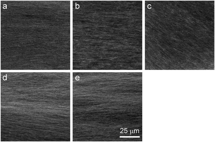
Microtubule organization in the axoplasm interior is well preserved after incubation with microtubule-targeting drugs. Axoplasm was incubated for 50 min in buffer alone (a) or in buffer containing 10 μM eribulin (b), 1 μM vincristine (c), 10 μM paclitaxel (d), or 10 μM ixabepilone (e), then fixed and processed for immunofluorescence microscopy with an anti-tubulin antibody (see Section 2). At this resolution, axoplasms incubated with drugs generally resembled untreated control axoplasms, with no evidence of extensive bundling or depolymerization observed. However, axoplasms with vincristine appear to have slightly more disorganized areas.