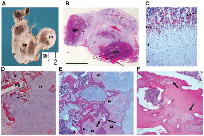
FIG. 5.
Morphology of the bones in metatropic dysplasia cases R08-023 and R09-035. A: Gross appearance of sectioned lower limb and histology of case R09-035. The femur and tibia are shortened with very little diaphyseal bone (arrow). There is extensive nodular overgrowth of disordered cartilage in the patella and at the ends of both long bones, which contain brown appearing bone marrow and bone. P, patella; DF, distal femur; PF, proximal femur; T, tibia. B: Low magnification of an H&E stained, paraffin-embedded whole section cut through the knee joint of case R09-035 and mounted on a 7.5 cm × 5 cm glass slide showing lack of growth plates, the nodular nature of the cartilaginous proliferation, and cortical bone (arrow) of the short femoral diaphyses. P, patella; F, femur; T, tibia; BM, bone marrow. C: Section showing the growth plate in case R09-035 illustrating the short hypertrophic zone and cartilage within the primary spongiosa (arrow). D: Growth plate from case R08-023 showing irregular primary spongiosa with trapped cartilage nodules (arrowhead) and a poorly organized hypertrophic zone (thin arrow). PS, primary spongiosa; H, hypertrophic zone. E: Nodules of cartilage (arrowheads) and woven bone (arrows) in case R09-035 arising in islands of mesenchymal cells. M, mesenchymal cell. F: H&E stained diaphyseal cortical bone from case R09-035 showing islands of immature woven bone (arrows) surrounded by normal lamellar bone.

