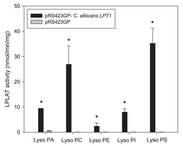
Fig. 1.

In vitro, microsomal LPLAT assays testing for C.a. LPT1 complementing S.c. lpt1Δ. LPLAT activity was measured in microsomes from S.c. lpt1Δ strains transformed with pRS423GP/C.a. LPT1 (4 CUG–UCG) or pRS423GP/– (vector only) 10 μg microsomes were incubated with 50 μM [14C]oleoyl CoA (20,000 dpm/nmol), 50 μM of lysoPA, lysoPC, lysoPE, lysoPI or lysoPS, and 100 mM Tris–HCl, pH 7.4 in 100 μl for 4 min. at 37 °C. After stopping the reactions, lipids were extracted, resolved using TLC, and quantified by scintillation counting. Data represent means ± standard deviation (n = 3) *p < 0.01.
